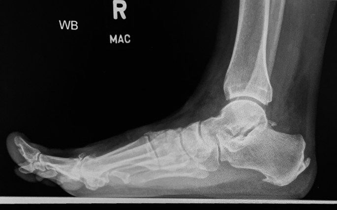
This is a Lateral view of a painful arch, Right foot.
image 4
“This is a radiograph of the right foot, Lateral view. With Achilles tendon insertion, and plantar fascia origin enthesophyte (spurring). There is a break in the anterior cyme line, and dorsal spurring of the 1st metatarsal head. The dorsiflexed toe shows limited range of motion.
My impression is… consistent with image 1, Hallux Rigidus with metatarsal head flattening, dorsal spurring, and decreased first metatarsal joint range of motion. Regnauld/Oloff classification Stage 3. ”
Answer: Hallux Rigidus
