
This is a AP view of a painful ankle, Left foot. In addition, here is a CT Coronal View of Talus.
Image 7
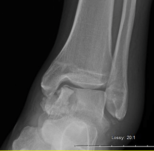
This is a radiograph of the AP view ankle as seen in image 1.
There is a sagittal sheer fracture of the medial talar dome, with associated comminution, and ankle joint dislocation. There appears to be a medial wall blow out of the lateral talus with decrease medial clear space. There is no sign of talar dome, calcaneal, or navicular bone fracture or dislocation. There is increase soft tissue volume and density of the medial ankle
The CT image shows significant communication and supports a sagittal sheer with associated transverse fracture patter of the talar dome.
My impression is a sagittal talar body fracture with communication, I will classify this as a Sneppen IIB.
